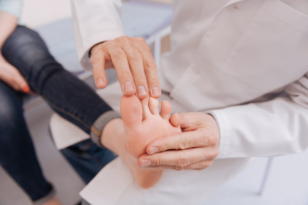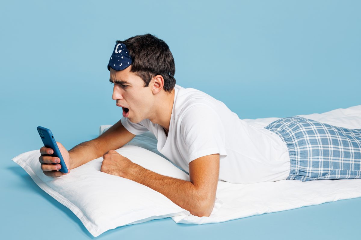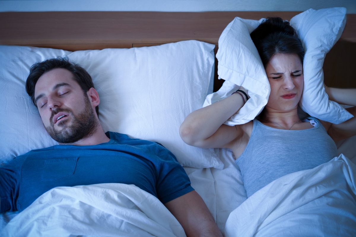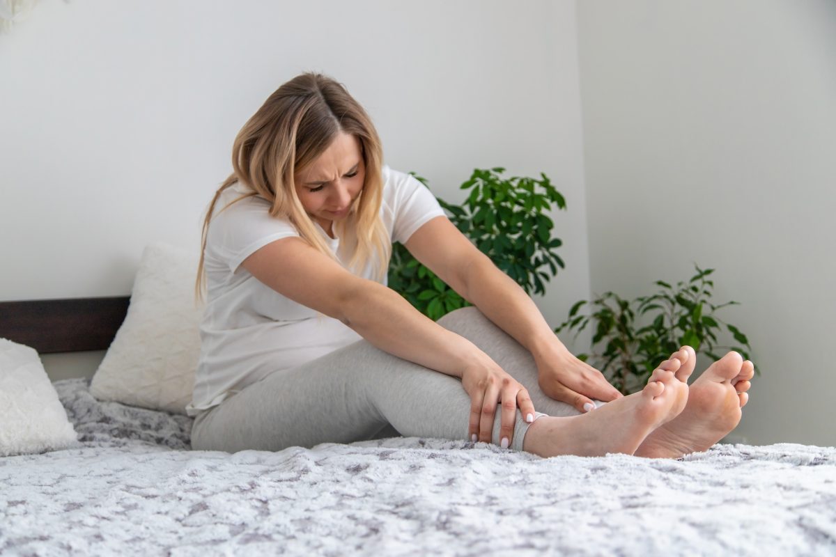
Your bedroom should be a place for rest, but for many people, calf cramps at night have made it a source of anxiety. That sharp jolt in your calf at 3 a.m. can leave you drained, unfocused, and uneasy for days.
The toll adds up far beyond the night itself; fatigue alone costs companies billions in lost productivity. Remarkably, all of this can begin with a single muscle spasm in the dark.
For years, the explanation seemed simple: dehydration or a lack of potassium. But that view overlooked what’s really happening.
Now, researchers are linking nighttime cramps to nerve misfires, essentially a glitch in the body’s wiring. Instead of a missing nutrient, the problem lies in how nerve signals travel between the brain and the muscles. When that communication falters, the muscle locks up, heart rate rises, and stress hormones surge, keeping the body from fully resting.
Stretching alone isn’t always enough. You deserve a routine that actually helps your nerves reset and your muscles relax before sleep. A focused five-minute approach works with your body’s natural systems to quiet irritation and ease tension, reducing those sudden midnight spasms. With a little consistency, better rest — and steadier mornings — can become the norm.
Motor Neurons, Electrical Storm, and Calf Cramps at Night
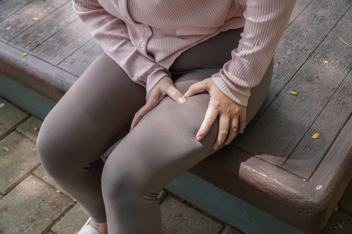
When it comes to calf cramps at night, know that your muscles are finely tuned engines powered by electrical signals. Within them are two small but essential sensors that keep everything in balance.
Muscle spindles monitor the length of each muscle, while tendon organs track the amount of tension being applied. Under normal conditions, these sensors communicate constantly, helping your body coordinate smooth, safe movement. When you stretch, the spindles signal the muscle to contract for protection, and the organs step in to ease that tension before it becomes strain.
Nighttime cramps happen when this communication system loses its balance. As you drift into sleep, your brain’s control over those muscle signals relaxes. If a muscle stays in a shortened or bent position too long, its sensors become overly sensitive — ready to react at the smallest trigger.
A minor shift under the covers can unleash a surge of nerve messages that tell the muscle to contract with full force. The result is sudden, intense pain and a muscle that feels locked in place, unwilling to release no matter how consciously you try.
The pathophysiology of myogenic muscle cramps, in contrast, is usually the result of disrupted energy production in muscle cells and occurs most commonly in metabolic myopathies associated with disorders of glycogen, lipid, or mitochondrial metabolism, according to Practical Neurology.
“Metabolic myopathies cause deficient ATP levels,” it states. “Because muscle relaxation is an adenosine triphosphate (ATP)-dependent active process, actin and myosin chains do not disengage, causing an electrically silent cramp (contracture). The metabolic defect may also cause accumulation of potentially toxic metabolites that further aggravate ATP deficiency. Myopathic cramps are also a potential symptom of myopathies linked to muscle membrane or intramuscular structural dysfunction in acquired and hereditary myopathies (muscular dystrophy, congenital myopathies, and inflammatory myopathy).”
Evolutionary Biology vs. Modern Bedding
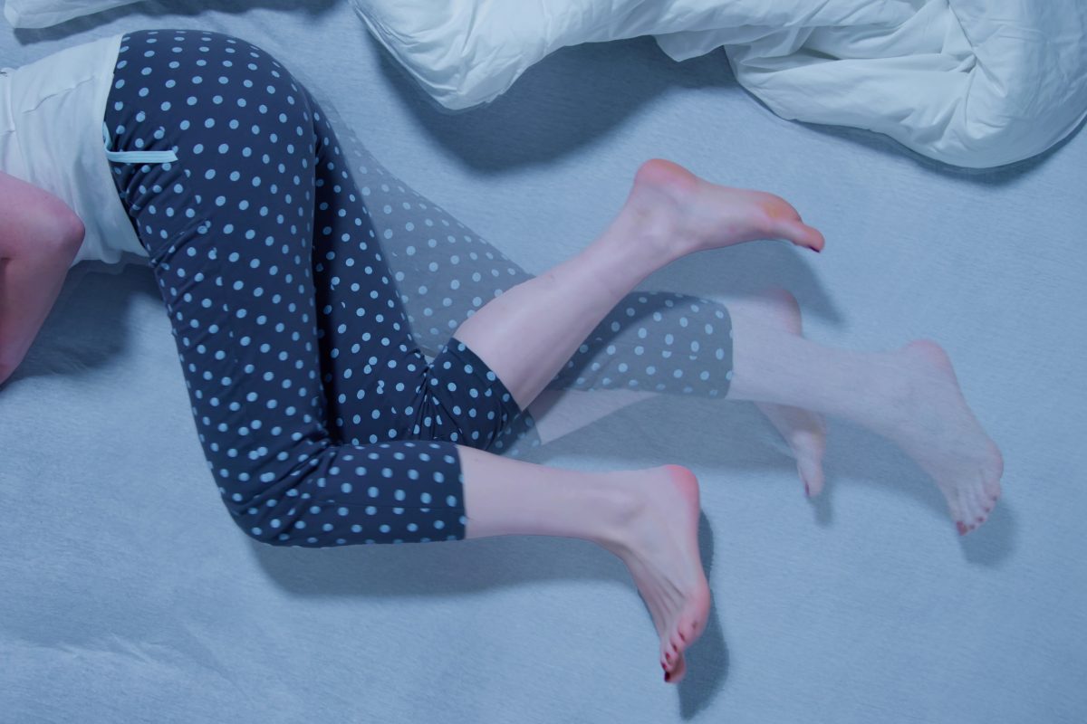
Plantar flexion is behind most calf cramps at night. Our bodies are designed to move best with the feet flat on the ground, but lying down changes that alignment.
Heavy or tightly tucked blankets often push the toes downward for hours, especially when you’re lying on your back. That constant pressure keeps the calf muscles shortened and tense, limiting blood flow and reducing the oxygen reaching those tissues.
Over time, those oxygen-deprived muscles become irritable at the cellular level. Ions begin to drift out of balance, disrupting the nerves that control relaxation. After several hours, even a slight movement can trigger a surge of activity, a deep and painful cramp that can jolt you awake in seconds.
The result is a sudden break in rest and a body that’s wide awake when it should be recovering.
“Occasional leg cramps are usually harmless, but it may be time to contact your health care provider if home remedies aren’t helping, your cramps are frequent or increasingly painful, they disrupt your sleep or daily activities, you experience muscle weakness, numbness or tingling in your legs, or you have related symptoms like back pain or unexplained swelling,” according to Banner Health. “Although nocturnal leg cramps can be painful, most people can find relief through stretching, hydration and lifestyle changes. And if these don’t work, medical care is available.”
90-Second Creep, Neurological Reset, and Calf Cramps at Night
Stretching often fails when you’re trying to get rid of calf cramps at night, because most people move too quickly. Short, jerky motions signal the body to protect itself by tightening the muscles, not relaxing them.
Longer, controlled holds are far more effective. Imagine a dry sponge — if you yank at it, it tears. But if you pull slowly and give it time, it softens. Muscles respond in much the same way.
To stretch properly, stand about a foot from a wall. Step one leg back and press your heel firmly into the ground, keeping your toes pointed forward. Hold this position for at least 90 seconds on each side.
Studies show it takes over a minute for muscle fibers and connective tissue to adapt and lengthen. At the halfway point, gently bend your back knee while keeping the heel grounded. This brings the soleus muscle into the stretch. The deeper calf muscle often responsible for persistent nighttime cramps.
Next, move to the edge of your bed. Sit with your legs extended and loop a towel or strap around the ball of one foot. Instead of pulling and holding, use a slow, rhythmic motion: pull the toes back and hold for about three seconds, then release for one.
Repeat this several times. This pumping action helps flush out stagnant blood and metabolic waste while drawing in fresh circulation. Your legs should feel lighter and more relaxed when you finish.
Once your muscles are loose, it’s time to calm the nerves that control them. The body cannot contract the calf and shin muscles at the same time. Instead, one set has to release when the other activates. This natural relationship is called reciprocal inhibition.
You can try a shin activation reset before bed. Lie down, pull your toes up forcefully toward your shins, and visualize the muscles on the front of your legs engaging. Hold for about ten seconds, and then release.
This movement triggers a relaxation signal from the spinal cord to the calves, something static stretching alone can’t achieve. Repeat this firm hard flex about five times. It quiets excess nerve activity and helps the lower legs settle down before sleep.
For the final step, perform a nerve slump. While seated, extend one leg, tuck your chin slightly, and alternate between pointing and flexing your toes. If you feel a light zing or stretch through the outer calf or foot, you’re on target.
Keep the motion gentle for 30 seconds. This small nerve glide helps reduce background electrical activity, which is one of the hidden triggers behind late-night cramps.
The Concrete Floor Syndrome and Rise of Sleep Ergonomics
Your middle-of-sleep calf cramps at night probably starts with what happens much earlier in the day. Workplace health teams were pointing to long hours on hard floors as a major driver of nighttime muscle cramps.
People who stand in one place for much of their shift develop increasing pressure and fluid buildup in their lower legs, which makes blood return to the heart more difficult. Over time, calves become tight, sore, and more likely to seize once you finally lie down to sleep.
Long stretches of sitting create a different but equally real problem. Remaining glued to a desk with your knees bent and your ankles still strains nerves behind the knee and reduces healthy muscle movement in the calves. Those muscles gradually stiffen, and the nerve pathways that control them become more reactive, setting the stage for cramps later in the night.
The solutions work best when they match the problem. If you spend much of the day on your feet, graduated compression socks can help move blood back up the leg. Look for socks labeled 20–30 mmHg that are snugger at the ankle and gradually looser toward the calf. This pressure gradient supports venous return and reduces that heavy, aching sensation by the end of the day.
If your workday is mostly seated, a simple ankle rocker or foot pedal under the desk can make a big difference. Moving your ankles through full flex-and-point pumps about 10 times every 30 minutes activates the calf muscles and veins, improving circulation without leaving your workstation. This small habit keeps the lower legs from shutting down and lowers the risk that tight, underused muscles will cramp once you fall asleep.
More recently, clinicians and sleep specialists have emphasized what some call ankle neutrality at night. This solution keeps your ankle in a natural, relaxed position instead of pulled sharply downward. With adjustable and hospital-style beds, you can achieve the optimal setup, where your knees and lower legs are slightly elevated to support blood flow and reduce strain on the calves.
For a standard bed, using two firm wedges usually works. One wedge goes under your knees and one under your calves. This is better than stacking loose pillows, which often collapse or shift.
Bedding tension matters, too. Tightly tucked sheets can pull the toes downward with several pounds of force, holding the ankles in a cramp-prone posture all night. A simple foot cradle or blanket-lifting frame keeps covers off the toes so the ankle can rest in a neutral position, reducing pressure on the calves.
If you sleep on your side, placing a firm pillow between your knees keeps the top leg from rolling forward, protecting the hips and lower back and decreasing the chance of waking up with extra soreness or leg discomfort.
Vascular Flow, Calf Cramps at Night, and Vitamin K2
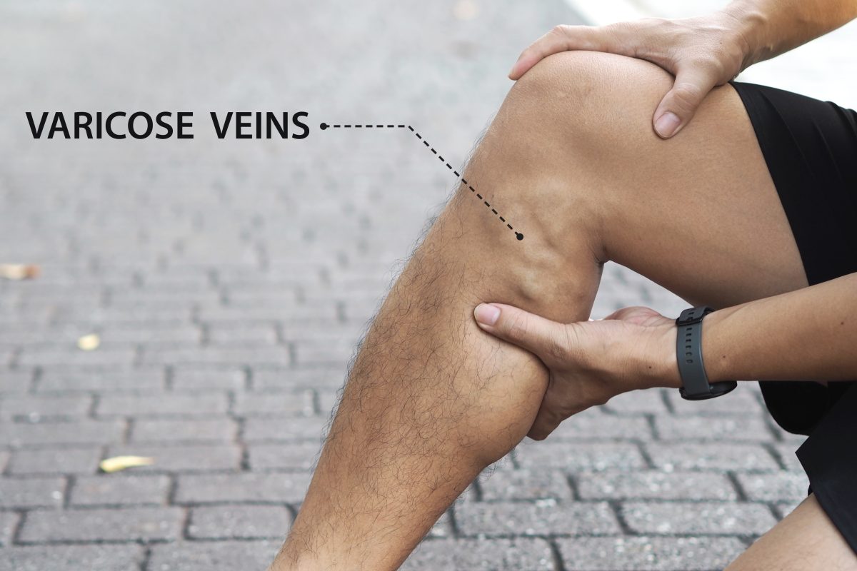
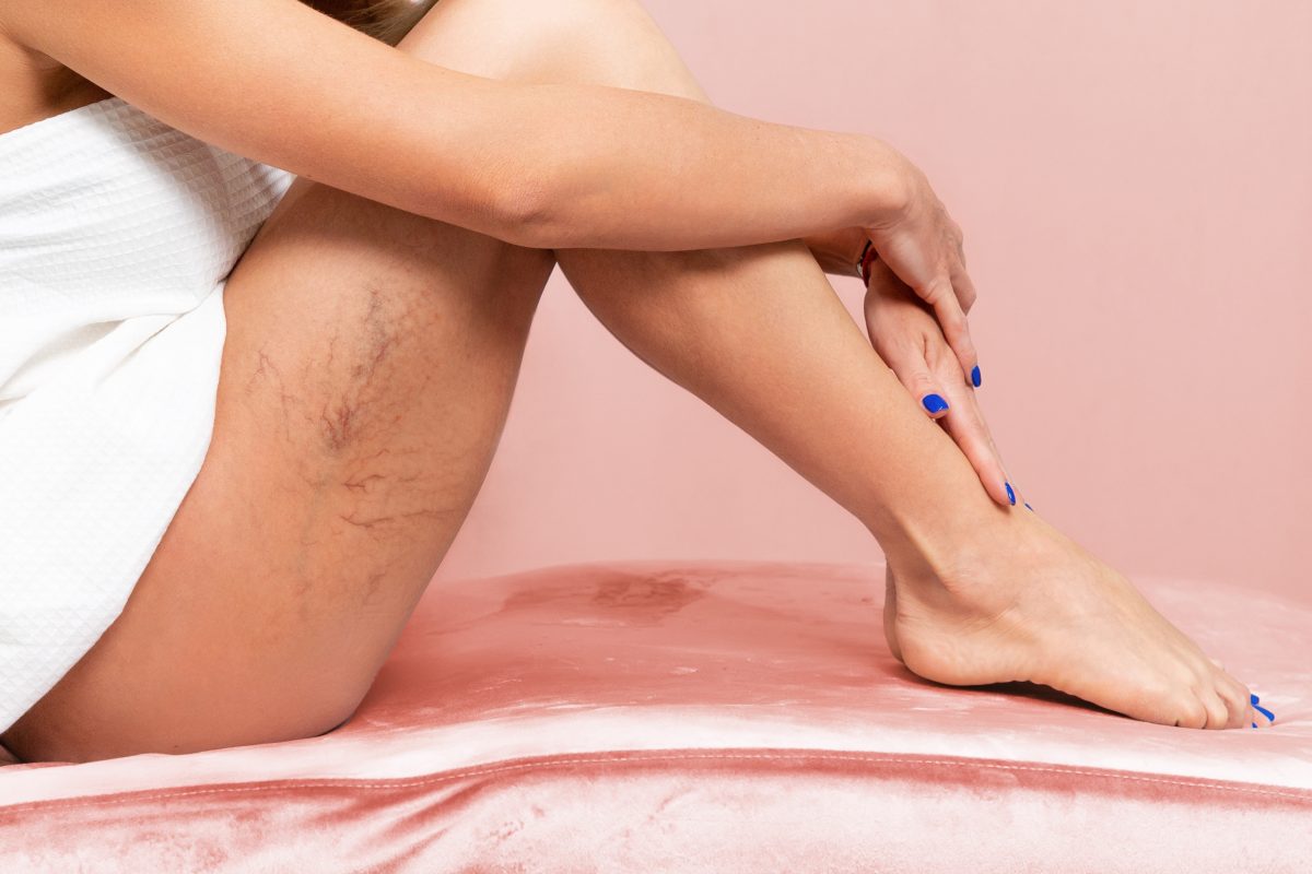
Those who feel they’ve tried every remedy for calf cramps at night may be facing something more complex than simple muscle weakness. In many cases, the underlying issue is how blood moves through the veins in the legs.
When the small valves inside those veins do not close properly, blood can pool and stagnate instead of flowing smoothly back toward the heart. Once you finally sit or lie down, this trapped fluid shifts, and the extra pressure around nearby nerves can disrupt how the muscles behave.
A focused treatment such as radiofrequency ablation can seal these faulty veins, usually easing nighttime cramps within days rather than months.
Additionally, standard nutrition advice often stops at magnesium, and that’s only part of the picture. Newer research has highlighted the role of Vitamin K2, particularly the MK-7 form, in keeping muscles able to fully relax. For a muscle to release, it must move excess calcium back out of the cell, and this process does not work efficiently when K2 is low.
Vitamin K2 activates proteins that help keep calcium from settling in the wrong places, including the muscles of the calves. In at least one large study, people who added K2 reduced the frequency of their cramps by more than half compared with those who relied on magnesium alone. With calcium better regulated, the muscle can completely relax.
| Intervention | Ideal For | 2026 Success Rate | Implementation | |
| Mechanical | 90-Second Fascial Creep | Beginners and active adults | Moderate (60%) | Zero Cost |
| Neurological | Reciprocal Inhibition | Chronic Advanced sufferers | High (85% Reset) | 2 Minutes |
| Ergonomic | Zero-G Incline / Wedge | Back sleepers and CVI patients | High (Prevention) | Home Investment |
| Nutritional | Vitamin K2 | Magnesium non-responders | High (Metabolic) | Supplement |
| Vascular | Vascular Ultrasound / RFA | Those who have tried everything | Very High (90% or More) | Clinical Visit |
| Tech | TOMAC Wearable | Neurological / RLS overlap | Emerging (High) | Prescription |
If stretching the muscle wall fails, try the neurological flex. If that doesn’t work, get a pain management specialist or doctor’s opinion on your blood flow.
Common Patient Questions
- Why do my cramps feel like a soreness hangover for two days after the event? A nocturnal cramp is a maximal voluntary contraction. You have essentially performed the equivalent of a 500-pound calf raise while you were asleep, which causes micro-tears in the muscle fibers. Most experts recommend light walking and heat.
- Can a stomach-sleeper ever truly avoid these cramps? It’s difficult because sleeping on your stomach forces your feet into 100-percent plantar flexion. If you cannot change your position, you must hang your feet off the end of the mattress. This allows your ankles to remain at a 90-degree angle.
- Is there a connection between cholesterol-lowering statins and my night cramps? Oftentimes, yes. Pharmacological reviews show that many statins can deplete CoQ10 levels in muscles. If you experience increased cramping, consider CoQ10 supplementation.
- Does pickle juice actually work for immediate relief? Surprisingly, for some patients, yes. Research shows the acetic acid in the vinegar triggers a reflex in the back of the throat that sends an immediate stop signal to the spinal cord.
- Should I wear socks to bed to keep my legs warm? Yes, if you choose the right ones. Loose, warm socks prevent thermal shock.
Teach Your Body: Nighttime is a Time to Unwind
Waking up with calf cramps at night can derail your entire night. Many people shrug them off, but anyone who has felt that sharp pull knows it’s impossible to ignore.
A focused five-minute routine can change that pattern so you move from simply coping to actively protecting your sleep.
Experts don’t know exactly what causes leg cramps at night, according to Healthline. “There are, however, known factors that can increase your risk,” the article states. “In most cases, nocturnal leg cramps are idiopathic, which means their exact cause isn’t known. Nighttime leg cramps may be related to foot position.”
It adds: “We often sleep with our feet and toes extending away from the rest of our bodies, a position called plantar flexion. This shortens the calf muscles, making them more susceptible to cramping.”
Instead of waiting for that sudden jolt, you can teach your body that night is a time to unwind. Muscles gradually soften, blood moves more freely, and your nervous system gets a clear signal that it’s safe to stand down.
Today, we know of tools like Vitamin K2 and targeted wearable devices that help fine-tune how the body handles muscle and nerve signals. Those advances matter, but the most meaningful shift often comes from what you do before you turn out the lights each night.
Lie flat, and take just over a minute to stretch your body in a controlled, steady way. Then add a few brief nerve-reset moves that calm the calves and feet. When these steps are combined, many people find they move from bracing for pain to drifting off more comfortably.
Try building this routine into your evening. Since your body has been signaling distress with every painful spasm, this is a practical way to finally respond.
Wellness and Pain
Find your personalized treatment for calf cramps at night by visiting Wellness and Pain. We offer conservative treatments, routine visits, and minimally invasive quick-recovery procedures. We can keep you free of problems by providing lifestyle education and home care advice.
This enables you to avoid and manage issues, quickly relieving your inhibiting lifestyle conditions when complications arise. We personalize patient care plans based on each patient’s condition and unique circumstances. Wellness and Pain can help improve wellness, increase mobility, relieve pain, and enhance your mental space and overall health.